


Next: Bias and overscan
Up:
The
ALFOSC Camera
Previous: Modulation Transfer Function
Quantum efficiency vs. wavelength:
With backside charging and a HfO coating, the QE reaches it's
maximum value of more than 90% from 450nm to 500nm, as can be seen
in the QE vs. wavelength plot in figure 5.
Shortwards of 400nm, the QE is decreasing, but remains relatively good,
with approx. 57% QE at 334nm.
Towards long wavelengths, QE decreases steadily due to the increasing
transmission of the Silicon of the thinned CCD. For observations
in the
  to
to  range,
one might try using a CCD temperature of
-80^
 C to increase the sensitivity slightly, but the dark current
will increase also and warm columns will appear.
Stability of the sensitivity:
In order to achieve high blue sensitivity, the Loral CCDs are thinned and
back side illuminated.
Still, short wavelength photons are absorbed near the back surface,
in a region where the potential from the electrodes is not felt.
To drive the photo-electrons towards the front side, charging
of the back side by UV-flooding was applied to CCDs previously used on the
ALFOSC. The charging proved sensitive to contamination, resulting in
unstable QE.
For this CCD, a Steward Observatory ``Cat-C'' coating is used.
The coating creates a potential difference at the Si surface
large enough to force the electrons to the front side.
In contrast to earlier coatings based on Platinum,
this coating has proved to be insensitive to contamination
from out-gassing when the liquid Nitrogen is used up.
Several global-scale QE measurements are displayed in figure
5. The QE is determined from the sigma-clipped average
ADU count of a 400 by 400 pixel area at the center of the CCD.
Some quite small changes in QE can be seen, at max.
The cause of this will be examined in the following.
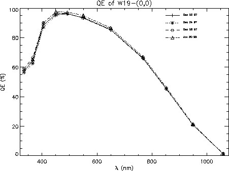
Figure 5:
Global quantum efficiency versus wavelength for the W19-(0,0) CCD,
measured from the sigma-clipped mean of the central area of a flat field
exposure.
Although recorded after highly different treatments, from UV-flooding to
a few days warm in poor vacuum, the QE is nearly unchanged.
Two curves are indistinguishable, dated December 02 and 04. In
between, the CCD was kept at -100^ C. This provides the ideal
environment for QE stability.
The January 30 graph shows slightly better QE over the entire
wavelength range. For this measurement, the strongly concave FOSC
entrance window was used, while for other measurements, a flat window
was used. The difference in QE results from a not quite perfect
compensation for the attenuation from the FOSC window. Also, another
controller was used, so another gain determination had to be used.
For this reason, the difference
between December and January data is not considered significant.
The December 02 and January 30 were both made immediately after
two days of storage at room temperature in the poor vaccuum from
out-gassing after the liquid Nitrogen tank runs dry. This has for
other coatings caused decrease of QE, but here it remains unchanged.
The change in local structure can be examined in figure
6 for 1060nm light, figure 7 for 550nm
and figure 8 for 334nm
With a few exceptions, the local structure is unchanged.
At all wavelengths, low-sensitivity specks are seen, the the QE of
some of these is not stable. Both increase and decrease of about 10% in
relative sensitivity can be found in the central area of a few of these
specks.
Some of the dark specks in the short wavelength flat field images are
apparently
caused by a locally too weak potential from the coating.
The potential can be increased in these regions by UV-flooding in an
Oxygen atmosphere. An example of the improvement in flat field uniformity
is shown in figure 9. Many of the small low-sensitivity
specks have disappeared. In figure 5, the December 05 measurement
has been made after UV-flooding. The global short wavelength QE appears
to have increased by about a percent after flooding. In fact, the
QE outside the specks is unchanged - the change in measured QE is only
due to the increased QE in the specks.
The stability of the UV flooding while the CCD is cold
has not been checked.
Previous experience with the lack of stability of UV-floodable CCDs
suggests that flooding to remove the specks is not worth the
effort.
Leaving the camera warm in vacuum for a few days completely removed
the effect of UV-flooding: the specks went back to the original
low sensitivity.
While the stability of QE is good under normal operation, the sensitivity
can be dramatically reduced under other circumstances.
After dismounting and re-installing the CCD into the camera,
a large area with low sensitivity appears, as shown in figure 10.
The sensitivity was seen to drop by as much as 1/3 of
the original level at 334nm.
The area was developed on two occasions, both showing exactly the same
structure, indicating the area is not defined by the distribution of
a contaminationg material, but is a property of the coating.
Some constituent of atmospheric air must be causing this, possibly water.
The QE is gradually re-gained when the CCD is in vaccuum at room temperature,
and if the CCD is heated to +50^ C for an hour while evacuating the
cryostat, the QE is completely restored afterwards.
Note that this problem does not occur even if the Nitrogen supply is
exhausted. Only direct exposure to atmospheric air has been seen to
cause problems.
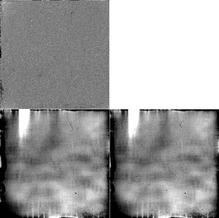
Figure 6:
Flat field properties at 1060nm.
Lower left:
The greyscale cuts are set to  of the median level.
The large scale structure with a peak to peak amplitude of about
10 %
directly relates to the thickness of the
CCD, almost completely transparent at this wavelength.
The vertical lines at the bottom is light reflected off electrodes
below the CCD.
The low sensitivity specks are relatively faint.
Lower right:
28 days later, the structure is essentially unchanged, although the CCD
has been UV-flooded, dismounted in atmospheric air, left warm in vacuum
and cold-cycled several times.
Upper left: Ratio between the two flat fields, displayed with
cuts of
  .
The large scale flat field structure is unchanged below the detection limit
in the central area, but near the edges, some changes of about 1% are seen.
In a few specks, the sensitivity has changed by
about 3%.
During entirely cold periods, QE changes have not been found.
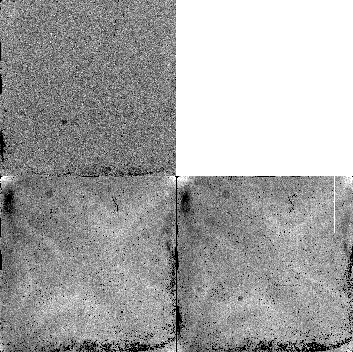
Figure 7:
Flat field properties at 550nm.
Lower left:
The greyscale cuts are set to  of the median level.
Uniformity is best around this wavelength.
Peak to peak large scale structure is about 1%.
Lower right:
28 days later, the structure is essentially unchanged, although the CCD
has been UV-flooded, dismounted in atmospheric air, left warm in vacuum
and cold-cycled several times.
Upper left: Ratio between the two flat fields, displayed with
cuts of
  . Except for a few displaced dust specks, the central large
scale flat field is unchanged to within 0.2%, but with 2% changes near
the edges.
In a few of the low sensitivity specks, the QE has changed by about 10%.
During entirely cold periods, QE changes have not been found.
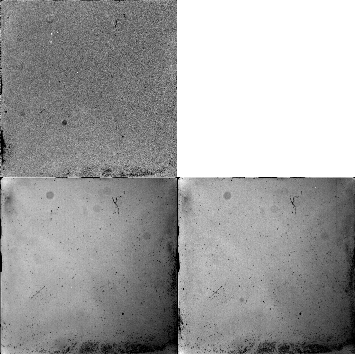
Figure 8:
Flat field properties at 334nm.
Lower left:
The greyscale cuts are set to  of the median level.
A 5% gradient is seen across the field.
Lower right:
28 days later, the structure is essentially unchanged, although the CCD
has been UV-flooded, dismounted in atmospheric air, left warm in vacuum
and cold-cycled several times.
Upper left: Ratio between the two flat fields, displayed with
cuts of
  .
The central large
scale flat field is unchanged, but 2% changes are found near
the edges.
In a few of the low sensitivity specks, the QE has changed by about 10%.
During entirely cold periods, QE changes have not been found.
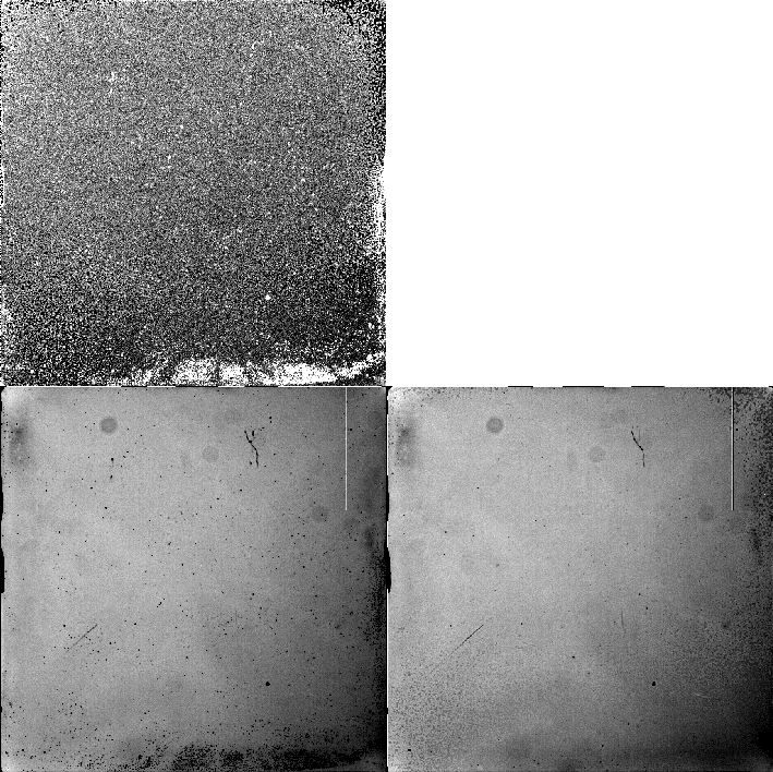
Figure 9:
Effect of UV-flooding for 334nm flatfields.
Lower left: Flat field before flooding.
The greyscale cuts are set to  of the median level.
Lower right: 3 hours after UV-flooding. Most of the low sensitivity
specks are gone.
Upper left: Ratio between the two flat fields, displayed with
cuts of
  . Areas with increased QE from UV-flooding appear bright.
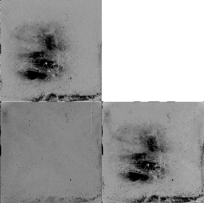
Figure 10:
Damage after exposure to atmospheric air:
Lower left: 550nm normal flat field.
Lower right: 550nm flat field made after the CCD was stored
in atmospheric air for a few days.
Upper left: Ratio between the two flat fields.
In the central region, the relative QE has decreased
by up to 35%.
All greyscale cuts are set to  of the median level.


Next: Bias and overscan
Up:
The
ALFOSC Camera
Previous: Modulation Transfer Function
Tim Abbott
Mon Apr 12 17:00:34 ACT 1999
 coating, the QE reaches it's
maximum value of more than 90% from 450nm to 500nm, as can be seen
in the QE vs. wavelength plot in figure 5.
Shortwards of 400nm, the QE is decreasing, but remains relatively good,
with approx. 57% QE at 334nm.
Towards long wavelengths, QE decreases steadily due to the increasing
transmission of the Silicon of the thinned CCD. For observations
in the
  to
to  range,
one might try using a CCD temperature of
-80^
 C to increase the sensitivity slightly, but the dark current
will increase also and warm columns will appear.

 C. This provides the ideal
environment for QE stability.
The January 30 graph shows slightly better QE over the entire
wavelength range. For this measurement, the strongly concave FOSC
entrance window was used, while for other measurements, a flat window
was used. The difference in QE results from a not quite perfect
compensation for the attenuation from the FOSC window. Also, another
controller was used, so another gain determination had to be used.
For this reason, the difference
between December and January data is not considered significant.
 C for an hour while evacuating the
cryostat, the QE is completely restored afterwards.
Note that this problem does not occur even if the Nitrogen supply is
exhausted. Only direct exposure to atmospheric air has been seen to
cause problems.

 of the median level.
The large scale structure with a peak to peak amplitude of about
10 %
directly relates to the thickness of the
CCD, almost completely transparent at this wavelength.
The vertical lines at the bottom is light reflected off electrodes
below the CCD.
The low sensitivity specks are relatively faint.
Lower right:
28 days later, the structure is essentially unchanged, although the CCD
has been UV-flooded, dismounted in atmospheric air, left warm in vacuum
and cold-cycled several times.
Upper left: Ratio between the two flat fields, displayed with
cuts of
  .
The large scale flat field structure is unchanged below the detection limit
in the central area, but near the edges, some changes of about 1% are seen.
In a few specks, the sensitivity has changed by
about 3%.
During entirely cold periods, QE changes have not been found.

 of the median level.
Uniformity is best around this wavelength.
Peak to peak large scale structure is about 1%.
Lower right:
28 days later, the structure is essentially unchanged, although the CCD
has been UV-flooded, dismounted in atmospheric air, left warm in vacuum
and cold-cycled several times.
Upper left: Ratio between the two flat fields, displayed with
cuts of
  . Except for a few displaced dust specks, the central large
scale flat field is unchanged to within 0.2%, but with 2% changes near
the edges.
In a few of the low sensitivity specks, the QE has changed by about 10%.
During entirely cold periods, QE changes have not been found.

 of the median level.
A 5% gradient is seen across the field.
Lower right:
28 days later, the structure is essentially unchanged, although the CCD
has been UV-flooded, dismounted in atmospheric air, left warm in vacuum
and cold-cycled several times.
Upper left: Ratio between the two flat fields, displayed with
cuts of
  .
The central large
scale flat field is unchanged, but 2% changes are found near
the edges.
In a few of the low sensitivity specks, the QE has changed by about 10%.
During entirely cold periods, QE changes have not been found.

 of the median level.
Lower right: 3 hours after UV-flooding. Most of the low sensitivity
specks are gone.
Upper left: Ratio between the two flat fields, displayed with
cuts of
  . Areas with increased QE from UV-flooding appear bright.

 of the median level.