 ions when in an Oxygen atmosphere, which causes
the back side to become negatively charged. The photo-electrons
generated near the surface of the CCD
are repelled and forced towards the electrodes
before recombination occurs.
In order to reach high quantum efficiency at short wavelengths, the
CCD is coated to create a ``passivated'' Platinum flash gate.
This adsorbs O ions when in an Oxygen atmosphere, which causes
the back side to become negatively charged. The photo-electrons
generated near the surface of the CCD
are repelled and forced towards the electrodes
before recombination occurs.
When the dewar becomes warm, outgassing of hydrocarbons occurs. These
combine with the O ions and cancel the negative surface charge,
leaving the CCD insensitive to short wavelength photons.
Procedure for Oxygen soaking:
After exposure to outgassing components or humid air, QE is restored in the following way:
Let the camera heat up to room temperature by setting the
CCD reference temperature to +20^\; C before the remaining
liquid Nitrogen
is used.
As strong outgassing
occurs, the camera should be pumped during the heating and
must be pumped for at least an hour when warm.
The camera is then filled with dry air to about ambient
pressure. In order to ensure no other air enters, a T-tube must
be connected to the dewar valve with the the other ends going to
the dry air bottle and a vacuum pump. If the pump is not equipped with
a valve, one must be inserted.
The tube between the the cryostat and bottle is then evacuated,
the valve to the cryostat opened, removing outgassing components from
the warm camera. The valve to the pump is closed, and dry air can now
slowly be let in while monitoring the camera pressure closely.
Fill the camera to slightly less than ambient pressure, then close
the cryostat valve.
Leave the camera with dry air at room temperature for at least an hour,
then evacuate and cool with LN
 .
The QE is now restored to specifications.
Quantum efficiency vs. wavelength:
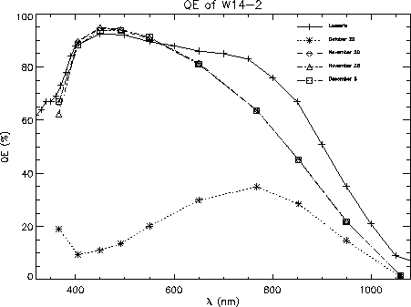
With backside charging and a HfO coating, the QE reaches it's
maximum value of approx. 90% from 400nm to 550nm, as can be seen
in the QE vs. wavelength plot in figure 5.
Within the uncertainties of the absolute calibration, the CUO and
Steward Observatory Lab measurements are in good agreement from 550nm
and shortwards, considering an uncertainty of about 10
CUO measurements.
At longer wavelengths, the measurements differ because of the temperature
of the CCD: room temperature for the SOL measurements and -100^\;
 C for
the CUO measurements. At high temperatures, the extra thermal energy
of the lattice makes the electrons easily excitable by low energy photons,
but the cost is a dramatically increased dark current. For observations
in the
  to
to  range,
one might try using a CCD temperature of
-80^\;
 C.
C.
Stability of the sensitivity:
Measurements performed at November 20 and 28 in figure 5 are both made 20 hours after Oxygen soaking and cooling. The identical curves show that a repeatable level is reached. 8 days after cooling the global QE remains at the same level, as shown by the December 6 graph. While the stability is excellent while the CCD is cold and in vacuum, the backside charge is completely destroyed by the outgassing that occurs at exhaustion of the liquid Nitrogen supply. This is illustrated by the October 23 graph, where the CCD was warmed up for one day and then cooled again without Oxygen soaking or pumping.

Figure:
Quantum efficiency of the W14-2 CCD.
Solid line with crosses is the measurement performed by the Steward Lab.
at room temperature.
The three almost coincident curves obtained at -100^\; C at the CUO lab.
shows the excellent stability when the camera is kept cold.
The lower curve shows the poor QE after ``Hydrogen poisoning''.
The measurements at 366nm have an uncertainty of about 10%.
In order to check whether the QE is still at the original level,
a blue beta-fluorescent source can be placed in the filter wheel
for a reference exposure.
On December 15 '96, a few hours after soaking and cooling, a level
of 57900 ADU was found in the central part of an image after bias
subtraction. The image was made with the settings: high gain,
amplifier A, binning 1 by 1.
The estimated temperature was +10^\; C. The source intensity has a
 temperature dependency, and also looses
  every year due to the radioactive decay.
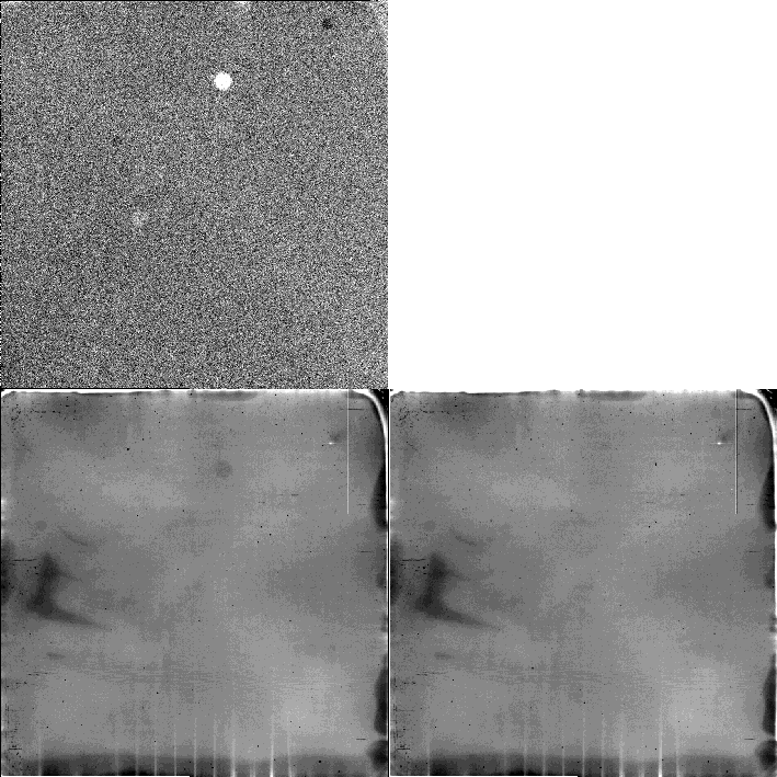
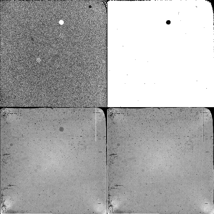
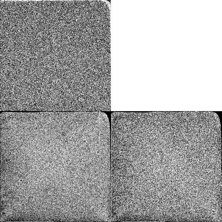
Flat field images at 1060nm, 550nm and 366nm illumination were obtained regularly during the cold period after soaking and cooling. Also local structure of the flat fields remains constant, as can be seen from the flat fields displayed in figures 6, 7 and 8. The pairs of flat fields are obtained 20 hours after and 8 days after cooling, and the ratio between the images sets an upper limit to the change in structure of 0.1% for the 1060nm and 550nm images and a limit of 1% at 366nm due to the lower signal to noise ratio. The only problem appears to be to keep dust away from the dewar window and filters!

Figure 6:
Flat field properties at 1060nm.
Lower left: 20 hours after cooling.
The greyscale cuts are set to  of the median level.
The large scale structure with a peak to peak amplitude of about
10 %
directly relates to the thickness of the
CCD, almost completely transparent at this wavelength.
The vertical lines at the bottom is light reflected off electrodes
below the CCD.
The low sensitivity specks are relatively faint.
Lower right: 8 days after cooling.
Upper left: Ratio between the two flat fields, displayed with
cuts of
  . Except for a few displaced dust specks, the flat
field is unchanged to within 0.1%

Figure 7:
Flat field properties at 550nm.
Lower left: 20 hours after cooling.
The greyscale cuts are set to  of the median level.
The ``X'' pattern extending from corner to corner is probably stray
light reflected off the rounded corners, and the central bulge may
also be due to stray light. Peak to peak large scale structure is
about 2%.
Lower right: 8 days after cooling.
Upper left: Ratio between the two flat fields, displayed with
cuts of
  . Except for a few displaced dust specks, the flat
field is unchanged to within 0.1%

Figure 8:
Flat field properties at 366nm.
Lower left: 20 hours after cooling.
The greyscale cuts are set to  of the median level.
Peak to peak large scale structure is
about 3%.
Lower right: 8 days after cooling.
Upper left: Ratio between the two flat fields, displayed with
cuts of
  . No changes with an amplitude of 1% or more are seen.